
Glucagon-like peptide-1, commonly referred to as glp-1, is an incretin hormone produced primarily in the enteroendocrine L-cells of the distal intestine and specific neuronal populations within the brainstem. First isolated in the 1980s following the cloning of the proglucagon gene, this metabolic peptide plays a foundational role in nutrient-induced insulin secretion and systemic energy homeostasis. Understanding its distinct biological mechanisms provides crucial insights into modern metabolic research and cellular signaling dynamics.
Endogenous Production and Metabolic Signaling Mechanisms of GLP-1
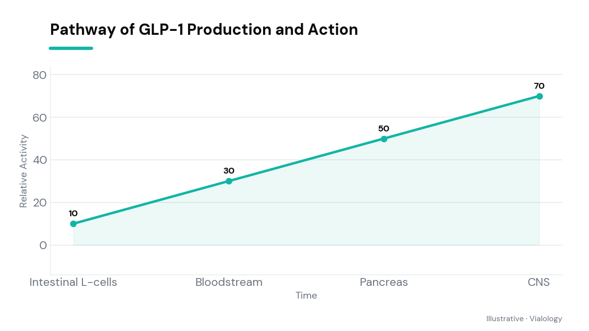
Endogenous synthesis of the glp-1 peptide begins with the tissue-specific enzymatic cleavage of proglucagon, a complex precursor prohormone encoded by the GCG gene. In response to nutrient ingestion—specifically dietary carbohydrates, long-chain fatty acids, and proteins—luminal enteroendocrine L-cells in the ileum and colon rapidly secrete active forms of the peptide, predominantly GLP-1 (7-36) amide, directly into the hepatic portal circulation. This meal-stimulated surge serves as a crucial physiological bridge between the gastrointestinal tract and systemic endocrine organs, initiating a coordinated cascade of metabolic responses designed to maintain postprandial glucose stability and optimize metabolic efficiency.
Once circulating, the hormone selectively binds to the GLP-1 receptor (GLP-1R), a classical class B1 G-protein-coupled receptor expressed across diverse tissues, including pancreatic beta cells, cardiac myocytes, vascular endothelial cells, and specific hypothalamic nuclei. Upon receptor activation, intracellular cyclic adenosine monophosphate (cAMP) levels rise sharply, triggering downstream protein kinase A (PKA) and exchange protein directly activated by cAMP (EPAC2) signaling pathways. In pancreatic islet cells, this intracellular cascade enhances glucose-dependent insulin exocytosis while concurrently suppressing glucagon release from neighboring alpha cells, thereby fine-tuning systemic blood glucose control without inducing hypoglycemia under normal physiological conditions.
Tracing the Research History of Incretin Biology and GLP-1
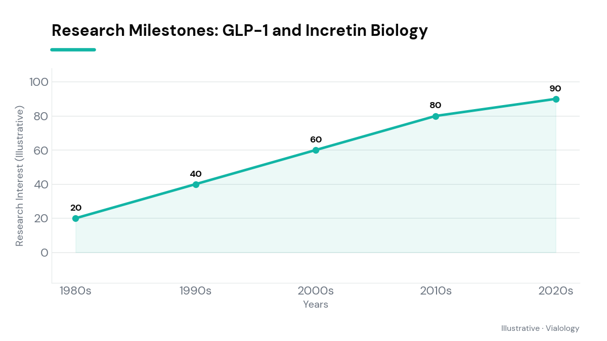
The scientific journey behind incretin biology dates back to the early 20th century, when researchers first hypothesized that intestinal extracts contained chemical messengers that stimulated internal pancreatic secretions during digestion. However, it was not until the mid-1980s that laboratory investigators successfully cloned the mammalian proglucagon gene and identified the specific amino acid sequences encoding GLP-1. Early in vitro and ex vivo experiments quickly demonstrated that truncated GLP-1 fragments, rather than the full-length peptide, possessed potent insulinotropic activity, inspiring a new wave of endocrine exploration focused on gut-brain metabolic crosstalk and peptide receptor kinetics.
During the 1990s, pioneering clinical research confirmed that continuous intravenous infusions of native GLP-1 significantly restored glucose sensitivity in human subjects with altered metabolic baselines. Despite these promising physiological findings, researchers encountered a severe pharmacological hurdle: native glp-1 exhibited an extraordinarily short biological half-life of less than two minutes in vivo. This rapid clearance driven by rapid enzymatic breakdown sparked dedicated structural engineering efforts to synthesize degradation-resistant receptor agonists, fundamentally reshaping modern peptide biochemistry and expanding the scope of target discovery in metabolic health.
Enzymatic Degradation and GLP-1 Pharmacokinetics
The rapid inactivation of native glp-1 in vivo is primarily mediated by dipeptidyl peptidase-4 (DPP-4), a ubiquitous serine protease expressed heavily on vascular endothelial surfaces, microvascular beds, and circulating in soluble form within plasma. DPP-4 rapidly cleaves the two N-terminal amino acids (Histidine-Alanine) from active GLP-1 (7-36) amide, converting it into the functionally inactive or weak antagonist metabolite GLP-1 (9-36) amide within seconds of secretion. This extraordinarily swift enzymatic degradation ensures that physiological incretin signals remain tightly regulated, brief, and locally localized to immediate dietary fluctuations rather than lingering unchecked in systemic circulation.
In addition to local DPP-4 cleavage, renal filtration and clearance represent a major secondary elimination pathway for circulating peptide fragments. The renal glomeruli efficiently filter both active and truncated forms of the peptide, which are subsequently reabsorbed and fully degraded by brush border peptidases within the proximal tubules. Because native peptides possess such transient systemic half-lives, modern biochemical research heavily emphasizes molecular modifications—such as fatty acid acylation, amino acid substitutions, or PEGylation—that sterically hinder DPP-4 binding while slowing renal filtration, thereby providing researchers with highly durable tools for studying metabolic pathways.
Central Nervous System Pathways and GLP-1 Satiety Regulation
Beyond its classical peripheral roles in pancreatic beta-cell function and gastrointestinal motility, glp-1 functions as a pivotal neuroendocrine transmitter within the central nervous system. Endogenous GLP-1 is synthesized directly by specialized preproglucagon (PPG) neurons located within the nucleus tractus solitarii (NTS) of the caudal brainstem. These central neuronal populations project widely to key autonomic and metabolic integration centers throughout the brain, including the arcuate nucleus, dorsomedial nucleus, and paraventricular nucleus of the hypothalamus, as well as mesolimbic reward structures such as the ventral tegmental area and nucleus accumbens.
Peripheral GLP-1 also signals to the central nervous system via vagal afferent sensory fibers residing in the hepatic portal vein and gastrointestinal submucosa, as well as through direct interaction with circumventricular structures that lack a dense blood-brain barrier, such as the area postrema. Activation of these central receptors suppresses hunger signaling, promotes postprandial satiety, and reduces reward-driven feeding behavior in extensive animal models. This integrated dual-pathway mechanism highlights how GLP-1 acts not merely as a simple digestive hormone, but as a master regulator connecting gut nutrient sensing to central energy balance.
Related on Vialology
Frequently asked questions
What is the biological role of natural GLP-1?
Natural GLP-1 acts as an endogenous incretin hormone that stimulates glucose-dependent insulin secretion, inhibits glucagon release, delays gastric emptying, and promotes satiety following nutrient ingestion.
How does GLP-1 interact with cellular receptors in research?
GLP-1 selectively binds to G-protein-coupled GLP-1 receptors on target cell membranes, elevating intracellular cAMP levels and triggering downstream signaling cascades such as PKA and EPAC2.
What did early clinical studies discover about GLP-1?
Early clinical trials in the 1980s and 1990s demonstrated that native GLP-1 infusions significantly improved postprandial blood glucose regulation in human subjects, though its physiological effects were extremely short-lived.
How is GLP-1 degraded in the human body?
Native GLP-1 is rapidly degraded within two minutes by the enzyme dipeptidyl peptidase-4 (DPP-4), which cleaves its N-terminal amino acids, followed by filtration and peptide breakdown in the kidneys.
Educational use only. Vialology publishes journalistic and educational content about peptide science. Nothing here is medical advice, diagnosis, or treatment, and nothing should be taken as an endorsement to use any substance. Many peptides discussed are experimental and are not approved by the FDA for human use. Always consult a licensed healthcare professional before making any health decision.